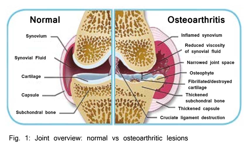
Osteoarthritis (OA) is considered one of the most important musculoskeletal disorders in both humans and horses. Clinically, it is associated with lameness and dysfunction of the affected joint and approximately 60% of all equine lameness is due to OA. It is characterised by breakdown and loss of joint cartilage, bony overgrowths (osteophytosis- seen on radiographs), stiffening of the fibrous joint capsule and its ligaments and synovial inflammation. Clinical symptoms therefore include pain and lameness, joint stiffness, swelling and fluid accumulation (joint effusion).

Anatomy of the Joint
Synovial joints are considered complex organs in which all constituent tissues (articular cartilage, subchondral bone, and synovial membrane) interact with each other, both directly and via the synovial fluid, in health and disease.
The joint capsule is the outer shell that protects the fluid filled cavity and its moving parts by attaching to the articular end of each bone involved in the joint. It is a sac-like envelope comprising fibrous tissue, tendons and ligaments that anchor it to the surrounding muscle and bone structures. The wall of the joint capsule is divided into an outer layer of fibrous tissue and an inner synovial layer.
Physiology of the Synovial Joint
The synovial layer is lined by a diverse population of synoviocytes, cells classified according to their ultrastructure. Type A cells are macrophages, implicated in phagocytosis of fluid, foreign material, and microbes. Type B cells are fibroblasts; locally derived cells that produce structural components including collagen. Type C cells appear to be an intermediate between type A and B forms. Beneath this synoviocyte cellular layer is the subintima, comprised of fibrous and adipose tissue, with blood vessels and nerves. The deepest layer is loose connective tissue that allows the membrane to move freely. Ligaments, tendons, or capsular fibrous tissue are located outside of that.
Two important molecules produced by synovial lining cells are lubricin and hyaluronic acid which help to protect and maintain the integrity of articular cartilage surfaces in synovial joints. Together, these two molecules reduce friction by providing boundary lubrication at the articular surface. As part of the OA complex, elastoviscosity of the synovial fluid is abnormally low.
What Causes Osteoarthritis
In athletic and young horses, synovitis and capsulitis are changes that occur early on and are assumed to be associated with repetitive trauma. This aetiology originates most often from overuse and conformational problems predisposing the horse to inappropriate biomechanical forces on the articular cartilage.
Multiple pathologic states develop after either single or repetitive traumas that are the initial “take-off” for progression towards OA. These can include traumatic injury, synovitis, capsulitis, sprain, intra-articular fractures, and meniscal tears. All of which lead to a common final end-stage of joint failure. Inflammation is most intense in acute synovitis and is one of the initial changes to occur in the development of OA Furthermore, the presence of synovitis in OA is associated with more severe pain, and joint dysfunction.This is shown to correlate with symptom severity, the rate of cartilage degeneration, and osteophytosis.
Refrences
- Goodrich, L.R. and Nixon, A.J., Medical treatment of osteoarthritis in the horse – A review. Vet J. 2006; 171: 51-69.
van Weeren, P.R. and de Grauw, J.C., Pain in osteoarthritis. Vet Clin Equine. 2010; 26: 619-642. - National Animal Health Monitoring Systems, Lameness and laminitis in US horses. Fort Collins, CO. USDA, APHIS, Veterinary Services-Centres for Epidemiology in Animal Health. 2000.
- McIlwraith, C.W., Principles and practices of joint disease treatment. In: Ross, M.W., Dyson, S.J., editors. Diagnosis and management of lameness in the horse. 2nd edition. Saunders. Missouri, 2011b; 840-852.
- Frisbie, D.D., McIlwraith, C.W., de Grauw, J.C., Synovial fluid and serum biomarkers. In: Joint disease in the horse. 2nd edition. Elselvier. St Loius, 2016; 10:179-191.
- Caron, J.P., Osteoarthritis. In: Ross, M.W., Dyson, S.J., editors. Diagnosis and management of lameness in the horse. 2nd edition. Saunders. Missouri, 2011; 655-668.
- Tnibar, A., Persson, A., Jensen, H.E., Svalastoga, E., Westrup, U., McEvoy, F., Evaluation of a polyacrylamide hydrogel in the treatment of induced osteoarthritis in a goat model: A pilot randomized controlled Study [abstract]. Osteoarthritis Cartilage. 2014; 22: 477.
- Tnibar, A., Schougaard, H., Camitz, L., Rasmussen, J., Koene, M., Jahn, W., Markussen, B., An international multi-centre prospective study on the efficacy of an intrarticular polyacrylamide hydrogel in horses with osteoarthritis: a 24 month follow up. Acta Vet Scand. 2015; 57: 20-27.
- McIlwraith, C.W., General pathobiology of the joint and response to injury. In: McIlwraith, C.W., Trotter, G.W., editors. Joint disease in the horse. Saunders. Philadelphia, 1996; 40-70.
- Brandt, K.D., Dieppe, P., Radin, E., Ethiopthogenesis of osteoarthritis. Med Clin North Am. 2009; 93: 1-24.
- Loeser, R.F., Goldring, S.R., Scanzello, C.R., Goldring, M.B., Osteoarthritis: a disease of the joint as an organ. Arthritis Rheum. 2012; 64: 1697-1707.
- Scanzello, C.R. and Goldring, S.R., The role of synovitis in osteoarthritis pathogenesis. Bone. 2012; 51(2): 249-257.